耳廓部分旋转在亚洲人除皱术中预防与矫正 pixie ear 畸形的临床观察
2026年10月25日25 min read
Aesthetic Surgery Journal
结论与实践要点
术式定位与适应证:本研究针对除皱术(扩大深平面除皱)后常见且具代表性的耳部畸形——pixie ear(文献报道发生率约 5.7%)提出「耳垂部分旋转」作为预防兼矫正手段。它适用于初次手术中为预防畸形者,也适用于既往除皱术后已出现耳垂畸形的二次修整;对于先天性耳垂附着异常者,其测量学标准亦可参考。需强调的是,本组为单一私人诊所的回顾性、非随机病例系列,证据等级有限。
关键技术点:A 组(n=40)采用 W 形切口联合 A 型肉毒毒素(BoNTA)切口周边注射;B 组(n=36)在此基础上于耳垂区做分段设计(45° 红段、直角橙/黄/白段),将黄段旋转 90° 后缝合于橙段、内层固定于耳前筋膜。术者可通过延长橙段、使橙-黄段成钝角来调节耳垂容积与游离尾段高度。关于发生机制的「皮肤过度切除引发牵引力、致耳前(垂直)与耳后(水平)提升矢量失衡」属于病理生理学推断,并非本研究的在体临床终点,其临床意义在于提示预防需兼顾设计而非单纯避免切除。
临床结局与并发症防范:76 例随访均值 12.5 个月(11–18 个月)。B 组术后 I-O/I-S 比值下降更显著(右耳 20.5%、左耳 22%),较A 组多改善约 8.4%/8.9%(P=.007/.006);四分箱显示B 组改善率大于 80% 且无一例恶化,A 组恶化率约 5%–15%。并发症方面无感染、不可逆神经损伤、严重不对称或涎瘘;但出现 5 例轻度皮肤坏死与 3 例暂时性下颌缘支麻痹,提示术区血供与神经保护仍须关注。瘢痕量表(MSS、MSRS、SBSES)两组无差异,说明增加旋转步骤未加重瘢痕。
证据等级与局限:本研究为回顾性、亚洲人群、术者单中心系列,未设随机对照,也未开展患者主观满意度调查(作者自承局限);照片测量可能受体位影响,且结论外推至其他人种需谨慎。对临床落地的建议是:该技法可作为除皱术的常规辅助步骤,但应以规范 W 形设计、精细缝合与避免皮瓣张力为前提,并据个体耳垂形态适度调整旋转量。
BackgroundThe most common deformity of the ear or earlobe appearing after a facelift is the pixie ear deformity.
ObjectivesThe purpose of this study was to devise and exercise a method to prevent ear deformity, including pixie ear, when performing a facelift and to correct it if it has already occurred.
MethodsExtended deep-plane rhytidectomy was performed between January 2017 and May 2024, and 76 patients who had passed 12 months post-surgery were included in this study. Group 1 consisted of 40 patients, who underwent W-plasty of incision and a combined botulinum neurotoxin type A (BoNTA) injection around the incision site. Group 2 consisted of 36 patients, who underwent rotation of the lower earlobe after W-plasty of the incision and combined BoNTA injection around the incision site.
ResultsIn a postoperative comparison between the 2 groups, Group 2 demonstrated superior outcomes over Group 1 by 8.4% on the right and 8.9% on the left, which was statistically significant ( P = .007, P = .006).
ConclusionsPartial rotation of the earlobe is demonstrated as a practical and effective surgical approach to prevent and correct pixie ear deformity.
Level of Evidence: 3 (Therapeutic)For image description, please refer to the figure legend and surrounding text.
Scars, sideburn distortion, lateral sweep, and deformities of the ear and earlobe are representative stigmata that reveal to others that a facelift has been performed after surgery and subsequent healing. Among these, there are some that cannot be avoided or are difficult to predict or prevent despite one's best efforts, but sideburn distortion or earlobe deformity can be improved with even minor efforts. 1
The most common deformity of the ear or earlobe appearing after a facelift is the pixie ear deformity. 2-7 Pixie ear deformity means that the cranial attachment segment increases, and conversely, the cranial free segment decreases. Furthermore, it refers to the loss or obliteration of the sulcus existing between the earlobe and the cheek's skin. The causes of occurrence are congenital cases and acquired cases because of distortion of the ear and earlobe caused by traction forces applied to the skin flap, scar retraction, and superficial musculoaponeurotic system (SMAS) flap. In fact, most acquired cases occur after a facelift, and even if efforts are made to eliminate the cause, the possibility of occurrence remains. 8 , 9
Therefore, a predictable, reliable, and definitive method is needed to prevent earlobe deformity while performing a facelift or to correct side effects already caused by a previous facelift. Accordingly, the purpose of this study was to devise and exercise a method to prevent ear deformity, including pixie ear, when performing a facelift and to correct it if it has already occurred. Having obtained good results, the author reports the research results and experience together with a literature review.
Extended deep-plane rhytidectomy was performed between January 2017 and May 2024, and 76 patients who had passed 12 months post-surgery were included in this study. Patients were divided into 2 groups. Group 1 consisted of 40 patients operated on until June 2022, who underwent W-plasty of incision and combined botulinum neurotoxin type A (BoNTA) injection around the incision site. After this combination, the author made changes to obtain better results. Group 2 consisted of 36 patients operated on after July 2022, who underwent rotation of the lower earlobe after W-plasty of the incision and combined BoNTA injection around the incision sit. Patients with a history of hypertrophic, keloid, or adverse scarring, use of systemic corticosteroids within the preceding year, recent heavy smoking, previous radiation treatment to the head or neck, or a history of rhytidectomy were excluded.
This study was conducted at a private clinic. The patients’ medical records and photographs were retrospectively reviewed. Formal institutional review board (IRB) approval (P01-202506-01-020, Public Institutional Bioethics Committee designated by the Ministry of Health and Welfare, Seoul, Republic of Korea) was obtained. All procedures performed in studies involving human participants were in accordance with the 1964 Declaration of Helsinki. Written consent was obtained from the patients, granting permission for the use and analysis of their data.
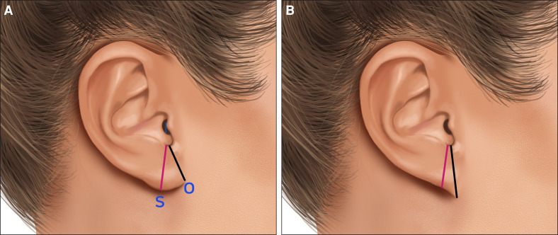
On the patient's lateral photograph, the distance from the intertragal notch (I) to the otobasion inferius (O, the lower-most point of the attached cephalic segment) was named I-O, and the distance from the intertragal notch (I) to the subaurale (S, the lowest point of the earlobe) was named I-S. Subsequently, each was measured and expressed as a ratio. This study also includes 25 congenital cases and 7 patients with pixie ears that occurred after a facelift. In such cases, the criterion for selecting the subaurale (S, the lowest point of the earlobe) was defined as the place where the tissue is thicker than other areas or where the contour of the lower earlobe changes abruptly ( Figure 1 ). An I-O/I-S value of 1 or less indicates normal, and 1 or more indicates a pixie ear. Also, to investigate the directionality of shape change before and after surgery, a 4-bin classification was performed. All measured values were classified into 4 sections (<0.8: excellent; 0.8 ≤ x < 0.9: good; 0.9 ≤ x < 1.0: fair; and ≥1.0: poor), and the rows represented the preoperative grade, the columns represented the postoperative grade, and the diagonal items represented no change (maintenance), improvement, and deterioration. In addition, a Sankey diagram was created to make it easy to recognize the changes before and after surgery at a glance.

Figure 1. Measurement method for a normal ear (A) and measurement method for a case of pixie ear deformity (B). The distance from the intertragal notch (I) to the otobasion inferius (O) was defined as I-O (black line), and the distance from the intertragal notch (I) to the subaurale (S) was defined as I-S (red line). Illustration created by Han Joon and Han Lyn.正常耳与 pixie ear 畸形的测量方法:定义耳间切迹(I)至耳屏下点(O)的 I-O 距与至耳垂下点(S)的 I-S 距;I-O/I-S 不大于 1 视为正常、不小于 1 提示 pixie ear。
For scar assessment, the Manchester Scar Scale (MSS), the Modified Scar Rating Scale (MSRS), and the Stony Brook Scar Evaluation Scale (SBSES) were used. 10-12 Photographs were taken before surgery and approximately 12 months after surgery. A handout was created using these 2 photographs, and 2 aesthetic surgeons performed independent and blinded photographic analysis. They were shown the handout and asked to record the score of the scar assessment. Afterward, the data were aggregated and analyzed.
Statistical analysis for this study was performed using Python 3.11. General and clinical characteristics of the participants were summarized using descriptive statistics. Continuous variables, such as age, preoperative and postoperative I-O/I-S ratios, and scar assessment scores, were characterized by mean and standard deviation. Categorical variables, including sex, history of rhytidectomy, and the etiology of pixie ears, were expressed as frequencies and percentages.
Changes in continuous variables within the same group before and after surgery were analyzed using paired t -tests, whereas independent t -tests were used to compare continuous variables between Group 1 and Group 2. Changes in the I-O/I-S ratio were analyzed separately for the right and left sides. The mean difference between preoperative and postoperative values was calculated for each group, along with 95% CIs. Additionally, Cohen's d was calculated to quantify the effect size of the surgical changes. All statistical analyses were 2-tailed, and a P -value of <.05 was considered statistically significant.
Extended deep-plane rhytidectomy with intravenous or general anesthesia was performed. After anesthesia, the design for surgery was made. It was designed in a small-sized zigzag pattern from the temporal area to the tragus. In Group 1, the zigzag pattern incision starts from the prehelical area and passes through the midpoint of the tragus ( Figure 2A ). 13 Then, the small zigzag pattern incision continues from the intertragal notch to the otobasion inferius. Once it passes the otobasion inferius and moves to the posterior auricular area, a medium-sized zigzag incision passes through the postauricular groove and reaches the occipital hairline. After all processes are completed, the remnant skin is trimmed in a zigzag pattern and sutured ( Figure 2B ). In Group 2, other areas except the earlobe are designed and incised in the same way as Group 1. On the other hand, in the earlobe, a 5 mm segment is drawn at a 45° angle anteriorly from the intertragal notch ( Figure 3A , red segment), and from there, a 1 cm segment is drawn at a right angle in a clockwise direction (orange segment). Then, a 5 mm segment is drawn at a right angle in a counter-clockwise direction (yellow segment), and a segment is drawn again at a right angle in a clockwise direction to the otobasion inferius (white segment). When passing the otobasion inferius and going up along the back of the earlobe, it ascends in a mirror-image form with the front. Then, before skin trimming and excision, the yellow segment of the pre- and postlobular area is rotated 90°, the skin is sutured to the orange segment, and the inner tissue is fixed to the preauricular fascia. Afterward, the remaining skin is trimmed, and the surgery is completed with the remaining skin suture ( Figure 3B , Video).
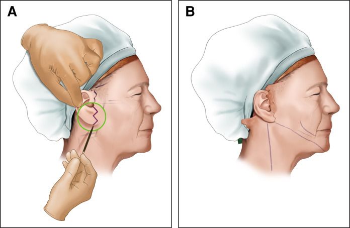
Figure 2. Preoperative design of Group 1 (A), State after suturing (B). Illustration created by Han Joon and Han Lyn.A 组(W 形切口联合 A 型肉毒毒素注射)的术前设计(A)与缝合后状态(B),耳垂区未作旋转处理,作对照基线。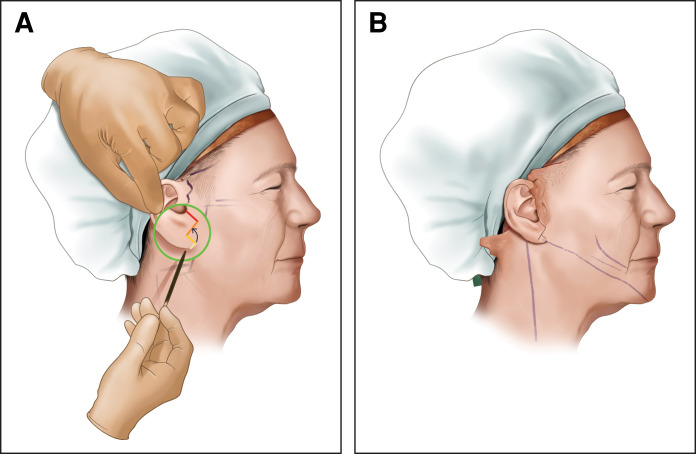
Figure 3. Preoperative design and process of rotating the yellow segment of the lobular area 90° of Group 2 (A) and state after finishing suturing (B). Illustration created by Han Joon and Han Lyn.B 组在 W 形切口基础上于耳垂区以 45°/直角分段设计,并将黄段旋转 90° 后缝合于橙段、内层固定于耳前筋膜(A),示缝合完成状态(B)。
A total of 76 patients underwent extended deep-plane rhytidectomy. The characteristics of the patients before surgery are summarized in Table 1 . The mean patient age was 49.6 years (range, 38-68 years). The mean follow-up period was 12.5 months (range, 11-18 months). There were no complications such as infection, permanent nerve damage, severe asymmetry, or sialocele during the follow-up period. However, there were 5 cases of minimal skin necrosis and 3 cases of temporary nerve paralysis of the mandibular branch.
Table 1. Patient Demographics
| Group 1 | Group 2 | P -value | |
|---|---|---|---|
| No. of patients (female/male) | 40 (40/0) | 36 (35/1) | — |
| Age (mean ± SD, years) | 50.8 (±5.7) | 48.2 (±3.5) | .232 |
| No. of rhytidectomy | |||
| Primary | 35 | 30 | — |
| Secondary | 5 | 6 | |
| Cause of pixie ear | |||
| Congenital | 13 | 12 | — |
| Acquired (trauma/after rhytidectomy) | 4 (0/4) | 3 (0/3) |
The preoperative and postoperative I-O/I-S of the 2 groups were classified. Before surgery, the <1.0 and ≥1.0 groups were the most numerous, and after surgery, most moved to the <0.8 group ( Table 2 ). Regarding the I-O/I-S ratio of the right ear, a comparison of Group 1's preoperative and postoperative results showed a 13.5% decrease from 0.978 ± 0.1 to 0.846 ± 0.14, which was statistically significant ( P < .001). In Group 2, there was a 20.5% decrease from 0.974 ± 0.03 to 0.775 ± 0.07, showing statistical significance ( P < .001; Table 3 ). Although there was no statistically significant difference between the 2 groups in the preoperative comparison, Group 2 demonstrated an 8.4% greater value than Group 1 on the right ear in the postoperative comparison, which was statistically significant ( P = .007). For the left ear, a comparison of Group 1's preoperative and postoperative results showed a 14.6% decrease from 0.983 ± 0.09 to 0.84 ± 0.14, which was statistically significant ( P < .001). In Group 2, there was a 22% decrease from 0.981 ± 0.02 to 0.766 ± 0.08, showing statistical significance ( P < .001). Although there was no statistically significant difference between the 2 groups in the preoperative comparison, Group 2 demonstrated an 8.9% greater value than Group 1 on the left ear in the postoperative comparison, which was statistically significant ( P = .006). A 4-bin classification was established to determine the directionality of changes before and after surgery ( Figure 4 ). In Group 1, the improvement rate was 52.5% for both the left and right sides, whereas the maintenance rates were 32.5% and 42.5%, and the deterioration rates were 15.0% and 5.0%, respectively. Conversely, in Group 2, the improvement rate was high at over 80% for both sides, with maintenance rates of 16.7% and 19.4%, respectively, and no reported cases of deterioration. In other words, improvement in condition was observed in nearly all patients in Group 2, demonstrating a distinctly higher pattern of improvement efficacy compared with Group 1. The Sankey diagram created to visualize these results also confirmed a shift toward better outcomes in Group 2 ( Figure 5 ).
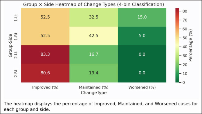
Figure 4. Four-bin classification created to investigate the directionality before and after surgery.按术前/术后 I-O/I-S 分级(优/良/中/差)的四分箱分类,直观呈现各组改善、维持与恶化的方向性变化。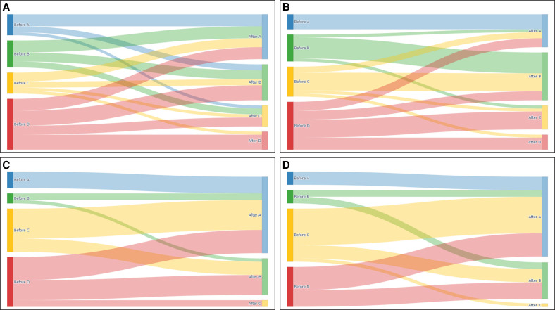
Figure 5. Sankey diagram to see the aspect of change before and after surgery (A: Group 1 left, B: Group 1 right, C: Group 2 left, D: Group 2 right).Sankey 图展示手术前后 I-O/I-S 分级流动(A 组1左、B 组1右、C 组2左、D 组2右),可见B 组多流向「优」级。Table 2. Grade Classification of I-O/I-S in Both Groups
| Group 1 | Group 2 | |
|---|---|---|
| Right | Left | Right |
| Pre | Post | Pre |
| <0.8 | 5 | 11 |
| <0.9 | 9 | 16 |
| <1.0 | 10 | 8 |
| ≥1.0 | 16 | 5 |
| Min | 0.57 | 0.45 |
| Max | 1.36 | 1.13 |
Table 3. Comparison of I-O/I-S Before and After Surgery Between Groups
| Preoperative | Postoperative | P -value* | Mean difference (95% CI) | d | |
|---|---|---|---|---|---|
| Right | |||||
| Group 1 | 0.978 ± 0.1 | 0.846 ± 0.14 | <.001 | 0.132 (0.087-0.177) | 0.94 |
| Group 2 | 0.974 ± 0.03 | 0.775 ± 0.07 | <.001 | 0.199 (0.155-0.244) | 1.52 |
| P -value | 0.778 | 0.007 | |||
| Left | |||||
| Group 1 | 0.983 ± 0.09 | 0.84 ± 0.14 | <.001 | 0.143 (0.095-0.192) | 0.94 |
| Group 2 | 0.981 ± 0.02 | 0.766 ± 0.08 | <.001 | 0.214 (0.178-0.250) | 2 |
| P -value** | 0.829 | 0.006 |
In the results regarding the MSS, the MSRS, and the SBSES for scar assessment, there was no statistical significance between groups in all 3 items ( Table 4 ). In other words, this indicated that even if rotation was performed, there was no difference in scarring compared with the control group.
Table 4. Comparison of Scars Between the 2 Groups
| Scale | Group 1 | Group 2 | P -value |
|---|---|---|---|
| Manchester Scar Scale(average score ± SD) | 5.42 ± 0.594 | 5.54 ± 0.628 | .409 |
| Modified Scar Rating Scale(average score ± SD) | 3.54 ± 0.486 | 3.5 ± 0.577 | .758 |
| Stony Brook Scar Evaluation Scale (average score ± SD) | 4.19 ± 0.539 | 4.24 ± 0.548 | .654 |
Pixie ear deformity means that the cranial attachment segment increases, and conversely, the cranial freesegment decreases. The causes of occurrence are congenital origin or acquired origin occurring after trauma, rhytidectomy, or total reconstruction of the ear, and most of the acquired origins are because of rhytidectomy. 2 , 4 , 7 It has been reported that the incidence of pixie ear deformity after rhytidectomy is 5.7%. 5 , 8 This figure accounts for a large proportion of the side effects caused by rhytidectomy. And another important reason is that it is a telltale sign. Therefore, efforts to reduce the frequency of such side effects must be made, and unlike other side effects, the frequency of occurrence can be significantly reduced with only minor effort ( Figures 6 - 8 ).
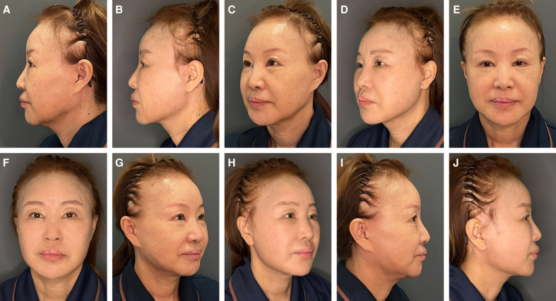
Figure 6. Photographs of a 60-year-old female patient who underwent secondary extended deep-plane rhytidectomy with the rotation of a lower part of the earlobe are presented. The preoperative images (A, left lateral; C, left oblique; E, frontal; G, right oblique; I, right lateral) and the postoperative images taken 12 months after the surgery (B, left lateral; D, left oblique; F, frontal; H, right oblique; J, right lateral) are shown.60 岁女性二次扩大深平面除皱联合耳垂下部旋转,术前(A/C/E/G/I)与术后 12 个月(B/D/F/H/J)各方位对照。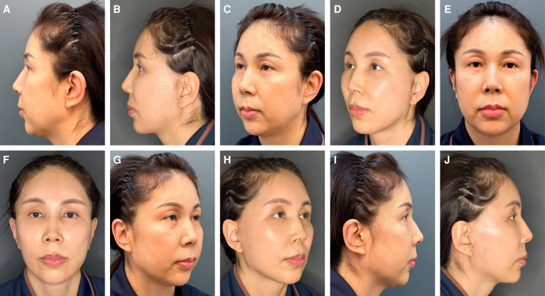
Figure 7. Photographs of a 54-year-old female patient who underwent primary extended deep-plane rhytidectomy with the rotation of a lower part of the earlobe are presented. The preoperative images (A, left lateral; C, left oblique; E, frontal; G, right oblique; I, right lateral) and the postoperative images taken 12 months after the surgery (B, left lateral; D, left oblique; F, frontal; H, right oblique; J, right lateral) are shown.54 岁女性初次扩大深平面除皱联合耳垂下部旋转,术前与术后 12 个月各方位对照。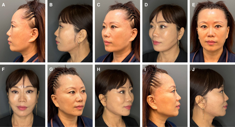
Figure 8. Photographs of a 48-year-old female patient who underwent tertiary extended deep-plane rhytidectomy with the rotation of a lower part of the earlobe are presented. The preoperative images (A, left lateral; C, left oblique; E, frontal; G, right oblique; I, right lateral) and the postoperative images taken 12 months after the surgery (B, left lateral; D, left oblique; F, frontal; H, right oblique; J, right lateral) are shown.48 岁女性三次扩大深平面除皱联合耳垂下部旋转,术前与术后 12 个月各方位对照。The fundamental cause of pixie ear deformity occurring after rhytidectomy is skin overresection. It is considered that this creates a strong traction force, which leads to hypertrophic scarring and an imbalance between preauricular (vertical) and postauricular (horizontal) skin-lift vectors. 9 Therefore, the first thing that must be preceded to not create a pixie ear is to avoid skin overresection. However, in the author's view, this alone cannot prevent side effects. This is because there were no problems in the early stages after surgery, but as time passes and more skin sagging occurs than expected, pixie ear deformity naturally follows.
Therefore, various surgical methods have been devised to correct or prevent pixie ear deformity. 14-24 These range from simple methods involving the removal of a portion of the earlobe skin to techniques that lift the entire ear using earlobe rotation flaps, skin redraping, or barbed sutures. The surgical method proposed by the author possesses several comparative advantages over other methods. First, existing surgical techniques are often limited to either the correction or the prevention of pixie ear deformity. 3 , 17-19 , 21 , 23 In contrast, although the author's technique primarily emphasizes prevention, it is also effective for correcting preexisting pixie ear deformities. Consequently, it is a viable routine procedure for rhytidectomy. Second, the surgical procedure is simple. Compared with traditional flap rotation methods, this technique is more intuitive and easier to master, allowing for immediate clinical application. 9 , 15 Third, It requires no additional procedures other than suturing following the preoperative design and incision. Furthermore, it avoids the need for the additional incisions and subsequent scarring often associated with conventional skin redraping or wedge resection methods. 22-24 Fourth, it is easy to adjust the earlobe's volume, curvature, or free caudal segment. This can be achieved during the preoperative design by elongating the orange segment and creating an obtuse angle between the orange and yellow segments. Doing so increases the volume of the earlobe to be resected and significantly elevates the lower part of the earlobe. Fifth, it does not negatively affect scarring. When compared with the author's previous research results, this method proved superior to the straight incision and showed no difference in results when compared with the control group (Group 1) where the same W-incision was applied. 13 Additionally, some may express concern regarding the potential formation of an additional scar (specifically where the orange and yellow segments overlap). However, there is no need for significant concern, because this area is neither conspicuous nor significant enough to impact the final outcome. Sixth, neither the occurrence nor recurrence of pixie ear deformity appears even after 1 year. Among the patients in Group 1 of this study, there were 6 cases where the pixie ear persisted because of insufficient correction on one or both sides, and 2 cases where it developed despite not being present previously. In contrast, there were no such cases in Group 2. The success rate was higher than those reported in previous studies. 15 , 23
However, disadvantages also exist. First, the operation time increases because of the additional suture and zigzag pattern skin trimming. Second, there is a possibility of wound dehiscence or delayed wound healing. Third, the ear might need to be pierced again. However, the disadvantages can be sufficiently overcome with little effort and meticulous suturing. In conclusion, the surgical method presented in this study can be routinely applied to all patients: for preventive purposes when there is no pixie ear deformity and for therapeutic purposes when pixie ear deformity is present.
This study also has limitations. First, because preoperative and postoperative photographs of patients were used when comparing results, there is a possibility that they were influenced by the patient's posture. Second, because the study was conducted on Asians, there may be differences when compared with other races. Third, a satisfaction survey to capture the patients’ subjective experiences was not conducted. Therefore, it is thought that studies with more patients, more items and over a longer period will be needed in the future.
Partial rotation of the earlobe appears to be a useful surgical method that may provide clinical value in preventing and correcting pixie ear deformity.
声明:中文精读 · 仅供学术参考。内容来自公开文献检索,不代表本人观点,不构成诊疗建议。 医疗美容需在正规医疗机构由执业医师实施。
基于相同主题推荐 · 继续深挖
本研究为韩国单中心回顾性系列,纳入 2017 年 1 月至 2024 年 5 月接受扩大深平面除皱、术后满 12 个月的 76 例患者,旨在预防并矫正 pixie ear 畸形。
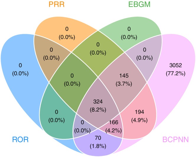
本研究利用美国 FDA 不良事件报告系统(FAERS,2004–2025)共 155,449 份 BoNT-A 相关不良反应报告,采用四种不相称性算法(ROR/PRR/BCPNN/
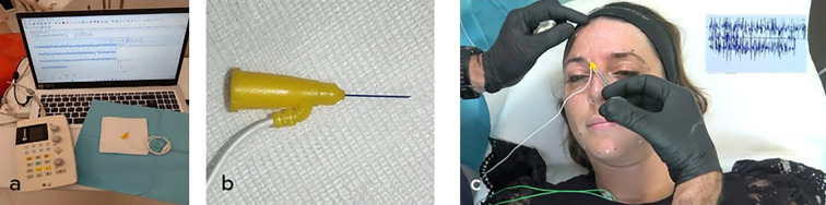
由6名意大利多学科专家(皮肤科、整形外科、神经科)基于文献回顾(PubMed 1973–2025)与临床经验形成的专家共识,针对incobotulinumtoxin A(Bocou